Abstract
Cost, quality and accessibility are major barriers to disease detection globally. For an easily communicable disease like tuberculosis, diagnostic or screening tests based on sputum, blood and urine analysis have slow response, are difficult to administer in remote locations, and have relatively high transportation and storage costs. Medical-grade state-of-the-art digital x-ray imaging systems are versatile in disease detection, faster, incorporate teleradiology for remote diagnosis, but are prohibitively expensive making them affordable only by major hospitals or labs that are located mostly in urban centers with high patient volumes. Here, a low-cost, high quality, digital x-ray imaging system could address many global health issues by enabling fast, accessible and inexpensive early detection of curable diseases including tuberculosis especially in rural, remote or under-populated areas.
In this research, we propose a path to achieving an inexpensive, high quality, digital X-ray system by focusing on the X-ray imager, a component that can reach 50% of the manufacturing cost of an imaging system. High manufacturing costs today are largely a function of small production volumes and various specialized fabrication processes. Our approach incorporates two technologies developed in-house that leverage existing manufacturing infrastructure because they are fully compatible with older generation amorphous silicon TFT display manufacturing lines: the first is a low dark current, high quantum efficiency optical radiation sensor that rivals state-of-the-art amorphous silicon PIN photodiodes and the other an amplified pixel circuit having a straightforward offset and gain correction scheme that yields higher signal-to-noise ratio than state-of-the-art passive pixels. When our sensor and pixel circuit technologies are integrated, the result is a high performance, low manufacturing cost diagnostic X-ray imager that can help achieve sustainable healthcare globally.
Biography
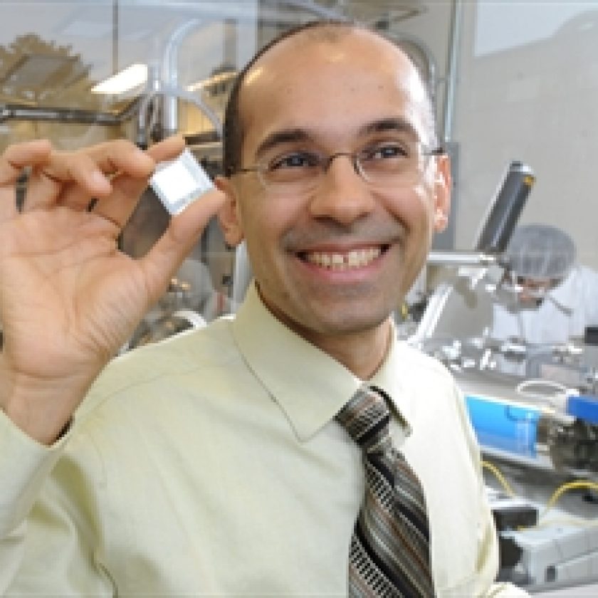
Karim S. Karim is currently a Professor in the Department of Electrical and Computer Engineering at the University of Waterloo. His research interests (http://star.uwaterloo.ca) encompass system, circuit, device and process development using amorphous semiconductors for digital imaging applications. Since 1998, he has co-authored 200 publications (mostly with his graduate students).
He is an IEEE Electron Devices Society Distinguished Lecturer, a Full Member of the American Association of Physicists in Medicine, and a registered Professional Engineer in Canada. Karim is also the Founder and CTO of KA Imaging, a medical device startup that designs and manufactures digital X-ray imagers for medical and industrial applications.


